IVF · Embryology · Andrology · Healthcare content
Embryology made simpler, one post at a time.
Hi, I'm Navya, and Embryopedia is my brainchild. I'm a former embryologist turned healthcare marketer — and I love explaining all things fertility, embryology and andrology, for patients and other embryologists (or just curious folks), via illustrations.

Worked with brands like


My story
From the IVF lab to marketing — to science illustrations.
I've spent time as an embryologist, working behind the scenes with eggs, sperm, and embryos under a microscope. But what I really loved was educating the patients who asked questions about their journey — and one day, I realised that I wanted to do this at scale. So, I switched to healthcare marketing, now helping thousands understand fertility and their journey better.
What started as cute science illustrations in my notebook has, four years later, grown into a full creative practice: a content brand, an audience of 4,500+ patients and clinicians, and millions of impressions on content that helps patients and embryologists alike.
Recent posts
Latest from the @embryopedia grid
Tap through the explainers — each one opens the post on Instagram.
Marketing for healthcare & femtech
I help healthcare brands reach the right audience — organically.
For four-plus years, I've been the content and organic-growth person for femtech and healthcare brands — bridging clinical accuracy with patient-friendly storytelling. Here's what I bring to the table: SEO and content strategy that brings you leads, topical authority that helps your brand rank, and copy and creative that improves brand recall.
Your turn
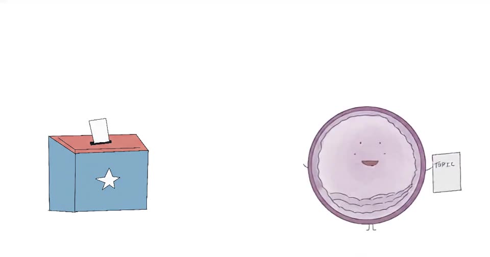
Have a topic you'd love me to cover next?
Drop it in the ballot box. Every Friday I pick one reader-submitted topic to turn into an Instagram post.
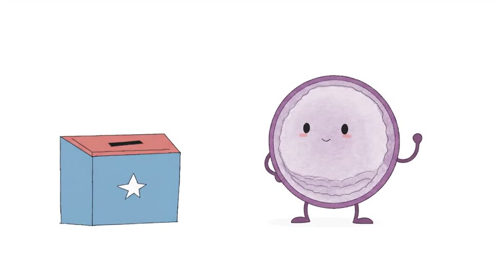
Got it! Your topic is in the box.
I'll let you know if it gets picked.